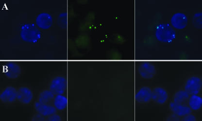
FIG. 3.
In vivo bactericidal activity of phage MSa. (A) Fluorescence microscopy pictures of kidney cells recovered from mice 4 days after intravenous infection with 108 CFU of GFP-expressing S. aureus. (B) Kidney cells from mice infected concurrently with 108 CFU of GFP-expressing S. aureus and 109 PFU of phage MSa. Cells were counterstained with 4′,6′-diamino-2-phenylindole (DAPI). (Left) Cells analyzed with a 340- to 380-nm filter (DAPI). (Center) Cells analyzed with a 450- to 490-nm filter (GFP). (Right) Overlay. Magnification, ×1,000 (oil immersion).

