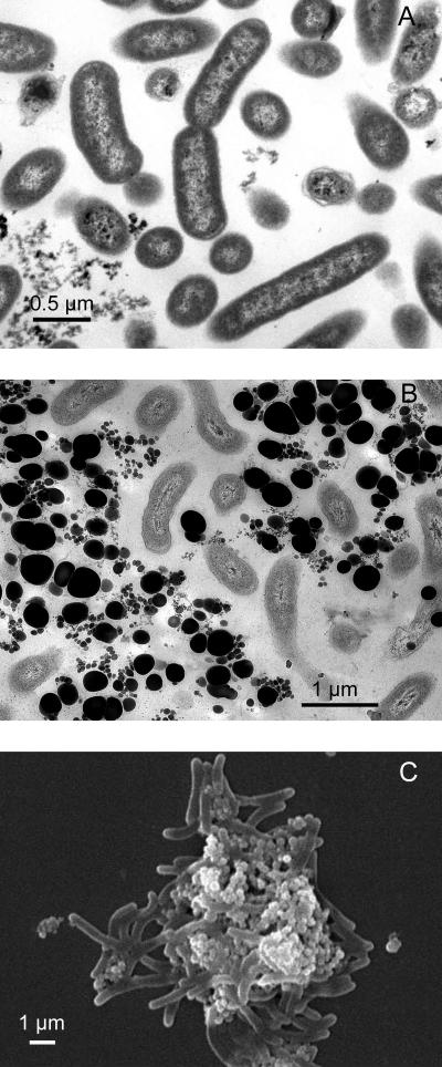
FIG. 6.
Electron micrographs of selenate-respiring isolate strain KM. (A) Transmission electron microscopy when the strain was grown without selenate. (B) Transmission electron microscopy when the strain was grown in the presence of selenate. (C) Scanning electron microscopy of cells grown in the presence of selenate.

