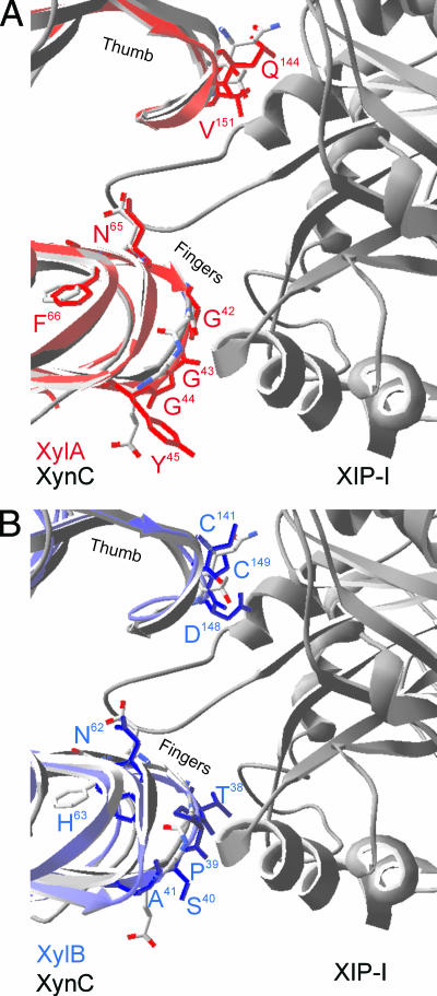
FIG. 1.
Molecular models of F. graminearum GH11 endoxylanases XylA (A) and XylB (B) superimposed upon the solved cocrystal structure of P. funiculosum XynC in complex with XIP-I. Ribbon representations of XynC (gray, left side), XIP-I (gray, right side), XylA (red), and XylB (blue) are shown. The “thumb” and “finger” regions, as well as side chains of putative specificity-determining residues, are indicated.

