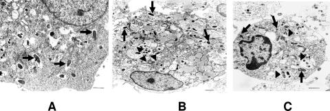
FIG. 1.
Ultrastructure of R. conorii-infected BMDCs and endothelial cells. Endothelial cells (A) and BMDCs derived from C3H mice (B) and B6 mice (C) were infected with bacteria (MOI of 5) for 24 h and then processed for ultrastructural analysis. (A) Numerous rickettsiae were detected in the cytosol of infected endothelial cells (arrows). (B and C) Bacteria were detected in both the vacuoles (arrowheads) and cytosol (arrows) of BMDCs. Data shown are representative images of three independent experiments with similar results. Bar, 1 μM.

