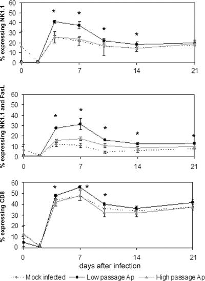
FIG. 3.
Total NK1.1, NK1.1/FasL, and CD8 expression in splenic lymphocytes was significantly higher in low-passage A. phagocytophilum-infected mice than in high-passage A. phagocytophilum-infected or mock-infected mice. Splenic lymphocytes were analyzed by flow cytometry using monoclonal antibodies to NK1.1 alone (top panel), to NK1.1 and FasL (middle panel), and to CD8 (bottom panel). Staining was performed individually, as described in Materials and Methods. The data are expressed as the proportion of splenic lymphocytes expressing the specific immunophenotype and are means ± standard deviations. An asterisk indicates that the P value is <0.05 for a comparison of low-passage A. phagocytophilum-infected mice and high-passage A. phagocytophilum-infected mice. Ap, A. phagocytophilum-infected mice.

