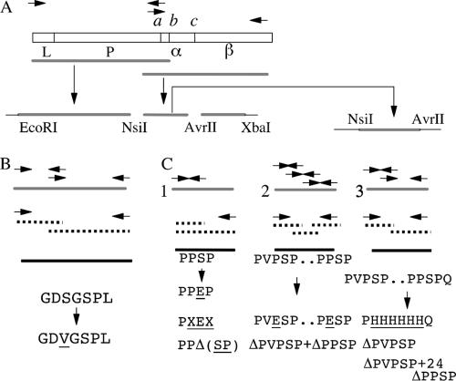
FIG. 1.
Cloning and mutagenesis of Neisseria meningitidis NMB IgA1 protease. (A) Physical map of a typical IgA1 protease gene and strategy involved in PCR amplification and cloning. The protease contains a signal peptide, L; a protease domain, P; cleavage sites a, b, and c (flanking the α-protein domain, α); and an autotransporter or β-core domain, β. (B) Schematic showing the approach used for site-directed mutagenesis of the active-site serine residue using the overlap extension method. (C) Site-directed mutagenesis of self-cleavage recognition sequences involved in IgA1 protease precursor autoprocessing using PCR-based megaprimer (1) and overlap extension (2 and 3) methods. Wild-type IgA1 protease sequence, solid gray line; PCR I products, broken line; PCR II products, thick black line; vector sequence, thick black line. PCR primers are represented by arrows. For details, see Materials and Methods.

