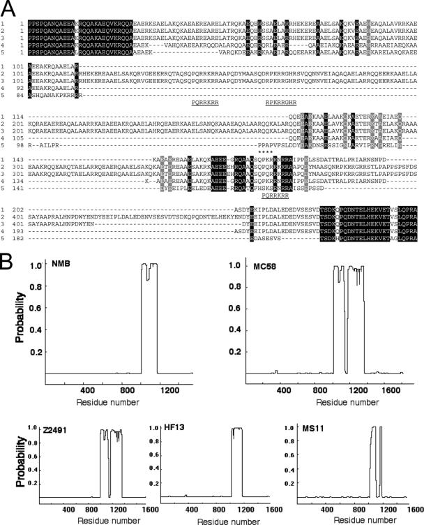
FIG. 2.
Sequence analysis of α-protein regions from N. meningitidis IgA1 protease sequences. (A) Multiple-sequence alignment of IgA1 protease sequences from N. meningitidis (1) NMB (AAK15023), (2) MC58 (NP-273742), (3) Z2491 (NP-283693), and (4) HF13 (CAA57857) and from (5) N. gonorrhoeae MS11 (P09790). The numbering of the α-proteins is relative to recognition site b (1003 in AAK15023). The position of the autoproteolytic site c in the MS11 sequence is also shown (marked by asterisks). Invariant amino acids sequences are highlighted in black and similar residues in gray. Putative nuclear localization sequences are shown underlined beneath the corresponding section of the alignment. (B) Coiled-coil prediction was carried out for the IgA1 proteases encoded by the indicated strains of N. meningitidis aligned in panel A using the Paircoil server (see Materials and Methods). The probability of each part of the amino acid sequences forming a coiled coil is represented.

