FIG. 4.
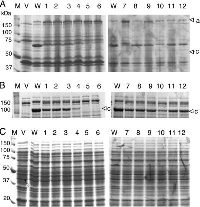
SDS-PAGE analysis of self-cleavage processing of wild-type and mutant IgA1 proteases of N. meningitidis NMB. (A) Accumulation of β-core fragments in outer membrane fractions as an indication of IgA1 protease self-cleavage activity in recombinant E. coli expressing IgA1 protease and its mutants. The upper arrowhead (a) denotes the position of the preprotein, while the lower arrowhead (b) shows the membrane-associated β-core fragment after self-cleavage. (B) Analysis of spent cell-free medium fractions concentrated 100 times. The arrowhead (c) denotes the mature processed IgA1 protease. (C) Analysis of cytoplasmic and periplasmic fractions. Lanes: M, molecular mass standards; V, E. coli BL21 host strain containing vector plasmid only; WT, clone producing wild-type IgA1 protease; 1 to 12, clones producing mutant variants of IgA1 protease enzyme. Table 1 shows details of mutant M1 to M12 sequences, and Fig. 3 shows quantitative determination of secreted IgA1 protease activity.

