Figure 2.
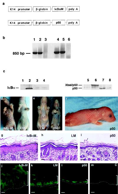
Transgenic mice engineered for gain and loss of epidermal NF-κB function. (a) Schematic of the K14-IκBαM and K14-p50 transgenes for targeted expression to murine epidermis. (b) Confirmation of transgene integration. Genomic DNA extracted from tail specimens of IκBαM[+] (lanes 1 and 2) and p50[+] (lanes 4 and 5) mice was subjected to PCR by using transgene-specific primers. Data obtained from unaffected littermates (lanes 3 and 6) with each primer set are also shown. (c) Western blot analysis of tissue protein extracts prepared from p50[+], IκBαM[+], and control mice. Lanes 1–4 are tissue extracts blotted with antibodies to IκBα. Lanes: 1, normal littermate skin; 2, IκBαM[+] skin; 3, normal littermate liver; 4, IκBαM[+] liver. Lanes 5–8 are blotted with antibodies to p50. Lanes: 5, normal littermate skin; 6, p50[+] skin; 7, normal littermate liver; 8, p50[+] liver. (d) Clinical appearance of K14-IκBαM transgenic mice. Five-day-old IκBαM[+] transgenic animals displayed clinical evidence of epidermal hyperplasia with markedly enhanced skin markings and a lack of visible hair growth compared with nontransgenic littermates. (e and f) Clinical appearance of 5-day-old p50[+] transgenic mice. p50[+] mice displayed clinical evidence of thin, slack skin compared with nontransgenic control. (g–i) Histology of IκBαM[+], age and site-matched control, and p50[+] mice; brackets define the thickness of the epidermis. Note increased epidermal thickness relative to normal in IκBαM[+] transgenic tissue compared with decreased epidermal thickness in p50[+] transgenic skin. (Bars = 75 μM.) (j–m) Epidermal expression pattern of p50 in (j) IκBαM[+] and (l) p50[+] transgenic mice along with (k) littermate and (m) secondary antibody alone controls.

