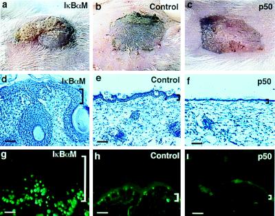
Figure 4.
Growth characteristics of murine epidermis transgenic for gain and loss of NF-κB function. (a–c) Clinical appearance of IκBαM[+], littermate control, and p50[+] skin harvested from 3-day-old transgenic mice and grafted to CB.17 scid/scid recipients, shown at 14 days postgrafting. (d–f) Histologic appearance of grafted skin from IκBαM[+], control, and p50[+] mice (scale bars = 150 μM). (g–i) The proportion of epithelial cells actively synthesizing DNA in vivo as a function of altered NF-κB activity. CB.17 scid/scid mice bearing IκBαM[+], p50[+], and nontransgenic control skin were injected with BrdU (250 mg/kg of body weight) i.p. Skin biopsy specimens were obtained 2 h later, and tissue sections were stained with antibody to BrdU. Shown are representative immunofluorescence micrographs. Note the increased labeling activity in IκBαM[+] skin that extends into cells of the suprabasal layers and the marked decrease in labeling activity in p50[+] skin compared with control. (Bars = 150 μM.)

