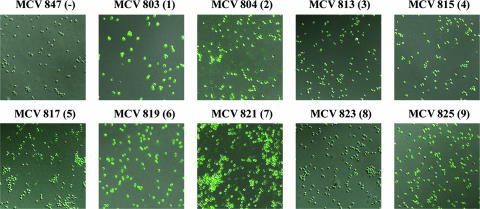
FIG. 2.
Immunofluorescence microscopy of TbpB-HA fusion strains. Iron-stressed gonococci were applied to glass slides and probed with an anti-HA monoclonal antibody followed by an Alexa 488-conjugated secondary antibody. Cells were visualized with a Zeiss LSM 510 Meta confocal microscope, at a magnification of ×63. Each image is labeled according to strain name with the HA epitope insertion number in parentheses. All TbpB-HA fusion strains are shown in a TbpA− background. MCV847 (TonB-HA fusion) serves as the negative control.

