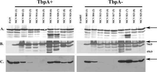
FIG. 3.
Expression of TbpB-HA fusion proteins. Iron-stressed gonococci were lysed, subjected to sodium dodecyl sulfate-polyacrylamide electrophoresis, and following transfer to nitrocellulose, probed with one of the following antibodies: polyclonal anti-TbpB serum (A) or peroxidase-conjugated high-affinity HA-specific antibody (B). Tf binding by the fusion proteins was assessed by probing with peroxidase-conjugated hTf (C). For both TbpA-expressing (+) and -nonexpressing (−) strains, each lane is labeled according to strain name with the HA epitope insertion number in parentheses. The arrows indicate the position of full-length TbpB (approximately 86 kDa). The positions of molecular mass markers are indicated on the right. FA19 and FA6905 serve as the wild-type and TbpB− controls, respectively.

