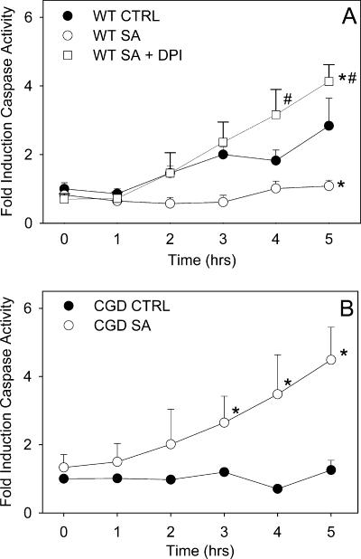
FIG. 5.
Caspase-3 activation in phagocytic murine neutrophils. Neutrophils were incubated either alone (CTRL) or with S. aureus (SA) (1:20) or were treated with DPI prior to coincubation with S. aureus (SA + DPI). Caspase-3 activity was assessed hourly by monitoring the increase in fluorescence with excitation at 390 nm and emission at 460 nm following cleavage of the fluorogenic peptide substrate DEVD-AMC. The means and standard errors of the means of 3 to 10 experiments are shown. An asterisk indicates that the P value is <0.05 for a comparison with the control, and a number sign indicates that the P value is <0.05 for a comparison with phagocytic neutrophils with a functional NADPH oxidase. WT, wild type.

