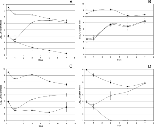
FIG. 6.
Colonization of the mouse intestine. (A) Sets of three mice were fed 105 CFU of E. coli MG1655 (+IS1) (▪), 105 CFU of MG1655 (+IS1) ΔflhD (▴), and 1010 CFU of E. coli MG1655 Δedd* (•). (B) Sets of three mice were fed 105 CFU of E. coli MG1655 (−IS1) (□), 105 CFU of MG1655 (+IS1) ΔflhD (▴), and 1010 CFU of E. coli MG1655 Δedd* (•). (C) Sets of three mice were fed 105 CFU of E. coli MG1655 (+IS1) (▪), 105 CFU of MG1655 (+IS1) ΔmotAB ΔfliC (○), and 1010 CFU of E. coli MG1655 (+IS1) Δedd* (•). (D) Sets of three mice were fed 105 CFU of E. coli MG1655 (−IS1) (□), 105 CFU of MG1655 (+IS1) ΔmotAB ΔfliC (○), and 1010 CFU of E. coli MG1655 (+IS1) Δedd* (•). At the indicated times, fecal samples were homogenized, diluted, and plated as described in Materials and Methods. Error bars representing standard errors of the log10 mean CFU per gram of feces for six mice are presented for each time point.

