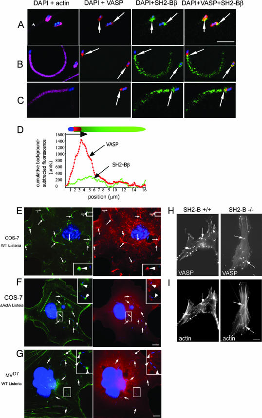
FIG. 6.
SH2-Bβ colocalizes with VASP and requires VASP for localization with actin. (A to C) COS-7 cells were infected with Listeria, stained with anti-SH2-Bβ (green), anti-VASP (red), phalloidin-Texas Red (pink), and DAPI (blue), and imaged by confocal microscopy. VASP colocalizes with SH2-Bβ on the side of the bacterium (A and B, yellow areas and arrows) and the beginning of the tail (B and C, yellow areas and arrows). *, the plasma membrane. Bar, 5 μm. (D) Cumulative fluorescence intensities for VASP (red) and SH2-Bβ (green) in the long actin tails were background subtracted and plotted as a function of position. (E and F) COS-7 cells overexpressing myc-SH2-Bβ were infected with either a WT (E) or ΔActA6 strain (F) of Listeria. Actin tails and actin caps at one bacterial pole were visualized by phalloidin-Alexa Fluor 488 (green, arrows), and myc-SH2-Bβ was visualized with an anti-myc antibody (red). Arrows from the images stained for actin and bacteria were superimposed on the images stained for myc-SH2-Bβ and bacteria and demonstrate that myc-SH2-Bβ localizes in the long actin tails and the actin caps formed by WT but not ΔActA Listeria. The boxed regions in the right corners are enlarged images of the marked areas in the larger images. Arrowheads from the images stained for actin and bacteria were superimposed on the images stained for SH2-Bβ and bacteria. Arrows with asterisks indicate actin caps formed by WT Listeria. Bar, 10 μm. (G) MVD7 fibroblasts were infected with WT Listeria. Actin caps were visualized by phalloidin-Alexa Fluor 488 (green, arrows), and myc-SH2-Bβ was visualized with anti-myc antibody (red). Arrows were superimposed as described above to demonstrate that there is no colocalization of these two proteins. The boxed regions in the upper right corners are enlarged images of the marked areas in the larger images. Arrowheads from the images stained for actin and bacteria were superimposed on the images stained for myc-SH2-Bβ and bacteria. Bar, 10 μm. (H and I) MEF from SH2-B+/+ (D) and SH2-B−/− (E) mice overexpressing GFP-VASP were infected with WT Listeria and stained for F-actin. GFP-VASP was localized at the poles of bacteria in both cell types. Arrows indicate actin tails (lower images) and GFP-VASP (upper images). Bar, 10 μm.

