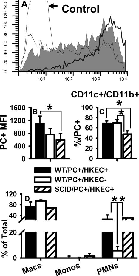
FIG. 9.
SCID pups treated with HKEC did not phagocytose DiO-labeled P. carinii as efficiently as did WT pups. SCID and WT pups were treated with aerosolized HKEC on days 4 and 2 previous to DiO-labeled P. carinii (PC) infection and on the day of infection. BALF was collected on day 1 postinfection. Cells were stained with antibodies specific for CD11c, CD11b, and MHC class II and examined for coexpression with DiO-labeled P. carinii. (A) Representative histograms showing DiO fluorescence of CD11c+ cells from HKEC-treated WT pups (thick black line), HKEC-treated SCID pups (medium line), and untreated WT pups (gray fill). (B) Mean fluorescence intensity (MFI) of DiO in CD11c+ cells. *, P < 0.05 compared to untreated WT and HKEC-treated SCID pups. (C) Percentage of CD11c+ CD11b+ cells positive for DiO; *, P < 0.05 compared to HKEC-treated SCID pups. (D) Differential counts of BALF cells; *, P < 0.05 compared to untreated WT pups. Data represent the means ± SD of four mice per group. Macs, macrophages; Monos, monocytes; PMNs, polymorphonuclear neutrophils.

