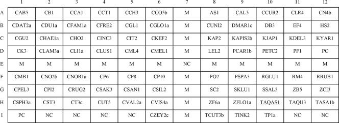
FIG. 1.
Layout of oligonucleotide probes on the array (0.75 by 0.9 cm). The positive control probe PC (located at D12 and I1) was designed from a conserved region of the fungal 5.8S rRNA gene. Probe NC (located at E7, I2 to I5, I11, and I12) was a negative control (tracking dye only). Probe M (located at E1 to E12 and at A7 to I7, except E7) was a DIG-labeled bacterial universal primer and was used as a position marker. The group-specific probe TAQAS1 (located at H10) is underlined. The corresponding sequences of all probes are listed in Table 2.

