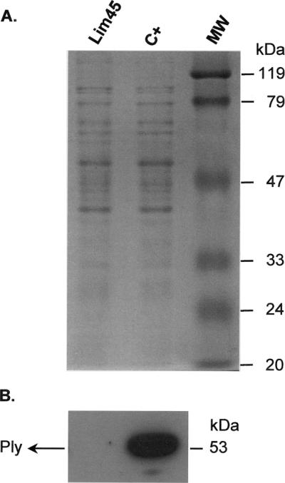
FIG. 1.
(A) Sodium dodecyl sulfate-polyacrylamide gel electrophoresis analysis of total protein from clinical pneumococcal isolate Lim45 (serotype I) and from a control (C+) pneumolysin-positive pneumococcal isolate. (B) Immunoblot analysis of Ply (pneumolysin) in clinical pneumococcal isolate Lim45 (serotype I) and in a control Ply-positive pneumococcal isolate. The membranes were probed with a rabbit antihuman-pneumolysin polyclonal primary antibody (1:5,000) and then incubated with a donkey anti-rabbit horseradish peroxidase-conjugated secondary antibody (Amersham Biosciences Europe GmbH, Vienna, Austria). Immunoreactive bands were detected with an enhanced chemiluminescence kit (Perbio Science Deutschland GmbH, Bonn, Germany). The position of the translational product in the control strain is indicated by Ply (53 kDa). MW, molecular mass.

