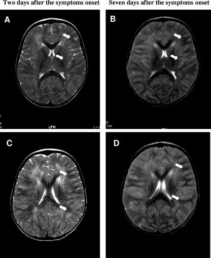
FIG. 1.
Brain MRI studies (axial, T2 weighted). (A and C) Axial, T2-weighted, diffuse, hyperintense signals with particular predominance in the periventricular and subcortical white matter of both cerebral hemispheres on day 2 after symptom onset (arrows). (B and D) Seven days after symptom onset, an axial, T2-weighted study revealed the extension of diffuse, hyperintense signals into the subcortical and deep white matter of both cerebral hemispheres (arrows).

