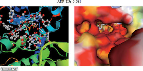
Figure 2.
An example of a view structure page. In the view structure page, interactive view of the putative complex is provided with two jV [15] panels. The complex structure with ribbon model (left panel) and that with the surface model (right panel) are presented. In the right panel, the residues within 5.0 Å from the putative ligand are specified with ball and stick model. These viewers can be rotated and translated with the mouse operation synchronously. In this example, the binding mode of ADP with the query protein (PDB: 1atp, E chain), predicted from the binding site appearing in 1l3r in PDB (c-AMP-dependent protein kinase), is shown.

