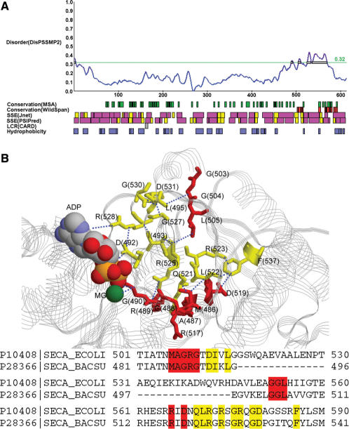
Figure 4.
(A) The result of P10408 (SECA_ECOLI). (B) The 10 residues predicted by both WildSpan and DisPSSMP2 are plotted as red sticks on the structure of a homologous protein, SECA_BACSU (P28366). Other 12 WildSpan predicted residues that either provides inter-molecular interactions to ADP or intra-molecular interactions with each other and/or the previous 10 residues are plotted as yellow sticks. Distances smaller than 5 Å are shown in blue dotted lines (PDB structure used: 1M74).

