FIG. 7.
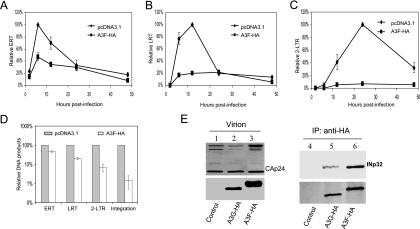
Effects of A3F on the early stages of HIV-1 replication during a single-round infection of HeLa cells. (A to D) HIV-1ΔVif was cotransfected with the pcDNA3.1 or A3F-HA expression vector and VSV-G into 293T cells. At 3 days posttransfection, HeLa cells were infected with HIV-1ΔVif that had been pretreated with DNase I. Total DNA was isolated from the infected cells at the indicated times following infection, and early RT, late RT, and 2-LTR circles were quantified. Virus input was normalized according to the level of p24. The highest value at peak productivity of HIV-1 from 293T cells cotransfected with pcDNA3.1 was set to 100%. (A) Quantification of early RT products. (B) Quantification of late RT products. (C) Quantification of 2-LTR circles. (D) Comparison of ERT, LRT, 2-LTR, and integration (Alu-based qRT-PCR) in the absence or presence of A3F-HA at the peak points. (E) HIV-1ΔVif was cotransfected with the pcDNA3.1, A3G-HA, or A3F-HA expression vector into 293T cells. At 48 h posttransfection, viruses were collected and purified from culture supernatants. Viral lysates were analyzed by immunoblotting using pooled HIV-1-positive human sera for the detection of viral proteins (lanes 1 to 3, upper panel) or an anti-HA MAb for the detection of virion-packaged A3G-HA or A3F-HA (lanes 1 to 3, lower panel). Viral lysates were also immunoprecipitated (IP) with the anti-HA MAb conjugated to agarose beads, and coprecipitated samples were then analyzed by immunoblotting using the anti-INp32 antibody (lanes 4 to 6, upper panel) or the anti-HA MAb to detect immunoprecipitated A3G-HA and A3F-HA (lanes 4 to 6, lower panel).

