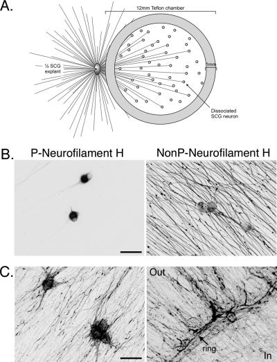
FIG. 1.
Isolator chamber culture system. (A) The system consists of a 12-mm Teflon chamber ring placed on top of the axons emanating from half of an SCG explant. Dissociated SCG neurons are plated inside the culture ring, contacting the axons from the explant. (B) Dissociated SCG neurons cultured inside the ring are mature after 1.5 weeks. Shown are confocal microscopy images of dissociated SCG neurons inside the ring fixed and stained for phosphorylated (P) neurofilament H (left panel) and nonphosphorylated (NonP) neurofilament H (right panel). In mature neurons, phosphorylated neurofilament H is restricted to the cell body and nonphosphorylated neurofilament H is restricted to axons. Scale bar, 50 μm. (C) Confocal images of dissociated SCG neurons inside the ring (left) and the edge of the ring (right) stained for F-actin with AlexaFluor 568-phalloidin. “Out” and “In” denote the outside and inside of the ring, respectively, and an arrow points out the ring border. Scale bar, 50 μm.

