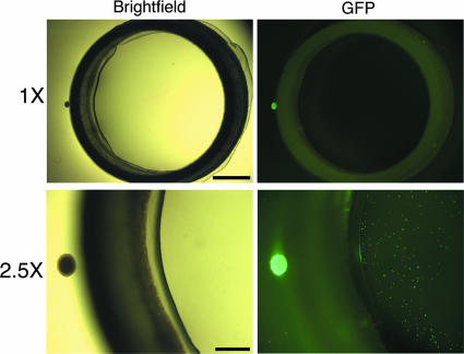
FIG. 2.
Anterograde transneuronal spread of PRV in the isolator chamber system. The explants were infected with PRV151, a recombinant expressing GFP, and fixed at 24 h postinfection. Scale bar, 1 mm. The top panels are 1× wide-field images of the isolator chamber system, showing the relative size of the SCG explant (small dot on left side of ring) and the Teflon ring. The bottom panels are 2.5× wide-field images showing the explant as a large GFP-positive dot to the left side of the ring and dissociated neurons inside the ring as small GFP-positive dots. Scale bar, 0.5 mm.

