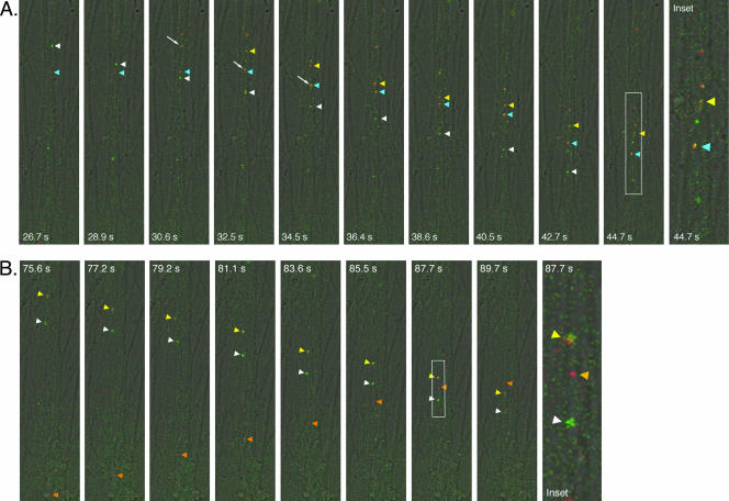
FIG. 8.
Live imaging of virion component dynamics within the isolator chamber using the Leica SP5 confocal microscope. The explant outside the ring was infected with PRV expressing a mRFP-capsid fusion and a gD-GFP fusion. The axons inside the ring run along the longitudinal axis of the frame and were imaged approximately 15.5 h postinfection. The ring edge and explant are located just beyond the top of the frame. The time scale shown at the bottom of each panel is synchronous with Movie S1 in the supplemental material. Figure insets are zoom images of the white outlined areas and refer to the time points indicated. (A). A gD-positive punctum (white arrowhead) crosses paths with a stationary punctum (blue arrowhead). The stationary particle “combines” with the yellow punctum (white arrow), and they proceed together. (B) A red fluorescent punctum (orange arrowhead) moves in the opposite direction from a gD-positive punctum and a yellow punctum and eventually crosses their path in the same axon.

