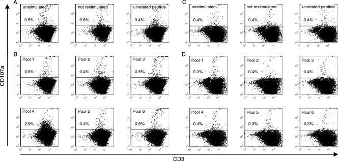
FIG. 2.
Representative dotplots of splenocytes from a woodchuck with resolved WHV infection (17214)(A and B) and from a naïve control animal (30341)(C and D). Lymphocytes were isolated and stimulated repeatedly in vitro for 6 days with WHcAg-derived peptide pools (B and D). As controls, cells were left unstimulated, not restimulated, or stimulated with an unrelated CMV peptide (A and C). Six animals that had resolved infection and two naïve control animals were analyzed for their responses to stimulation with peptide pools.

