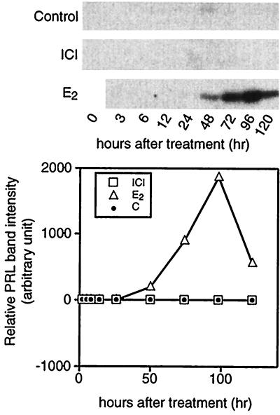
Figure 6.
Time course of intracellular PRL accumulation in PR1 cells cultured under different conditions. On the indicated day the cells were harvested and proteins were extracted by RIPA buffer as described. Equal amounts of protein were loaded in SDS/10% PAGE and Western blot analysis was performed to detect PRL bands. Each band was quantitated by densitometer. One nanomole of E2 or ICI was used.

