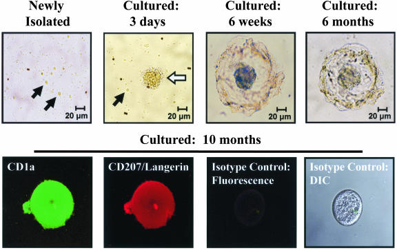
FIG. 1.
Differentiation of blood-derived pre-LC into LC during prolonged culture. Examination by light microscopy revealed that most of the CD1a+ pre-LC newly isolated from blood were small round cells (black arrows). After 3 days, the number of small round cells decreased and larger cell clusters with short dendrite-like projections (white arrow) appeared. By 6 weeks, most cell clusters elaborated a large spherical structure of interlocking dendritic projections surrounding the central core. These structures were stable for more than 10 months of culture and did not increase in number over time, suggesting a lack of cell division. Fluorescent immunostaining and imaging with a laser scanning confocal microscope demonstrated expression of both CD1a and CD207/Langerin in these cell clusters, confirming their differentiation into the LC phenotype. Staining with isotype control fluorescent antibodies was negative. DIC, Nomarski differential interference contrast.

