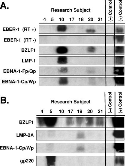
FIG. 5.
EBV M-RT-PCR of LC isolated from oral epithelium. CD1a+ LC were isolated from cells obtained by brush biopsy of grossly normal oral mucosal epithelia of seven healthy subjects. Half of each oral LC specimen was studied as newly isolated cells, and the other half was studied after culture of the cells in the presence of chemical inducers of EBV replication for 3 days. RNA extracted from the cells was studied by EBV M-RT-PCR amplification and specific probe hybridization. (+) Control = B958 lymphoblastoid cell line DNA and Akata Burkitt's lymphoma cell line DNA; (−) Control = no DNA. (A) Newly isolated oral LC. EBER-1, BZLF1, EBNA-1-Fp/Qp, and EBNA-1-Cp/Wp expression was demonstrated in subjects 10 and 20. LMP-1 expression was demonstrated in subject 10. LMP-2A and gp220 expression was not detected. RT +, with reverse transcriptase; RT −, without reverse transcriptase. (B) Cultured and induced oral LC. Newly induced BZLF1 expression was demonstrated in subjects 4, 5, 17, 18, and 21 and was especially strong in subject 5. (Note that the PCR product of subject 10 leaked from the well of the gel, explaining the apparent lack of BZLF1 hybridization for the induced cells of that subject, and that BZLF1 expression was also previously detected in subject 20 prior to induction.) Newly induced LMP-2A and EBNA-1-Cp/Wp expression was demonstrated in subjects 5 and 18. Newly induced strong gp220 expression was demonstrated in subject 5. LMP-1 and EBNA-1-Fp/Qp expression was not detected.

