FIG. 6.
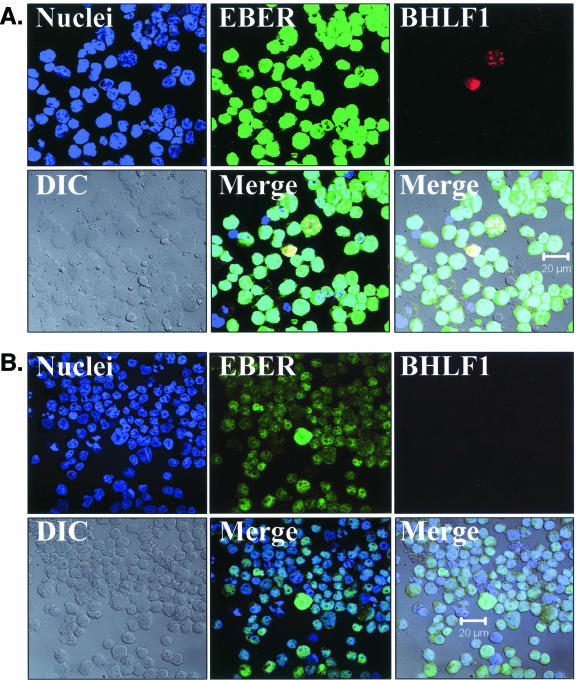
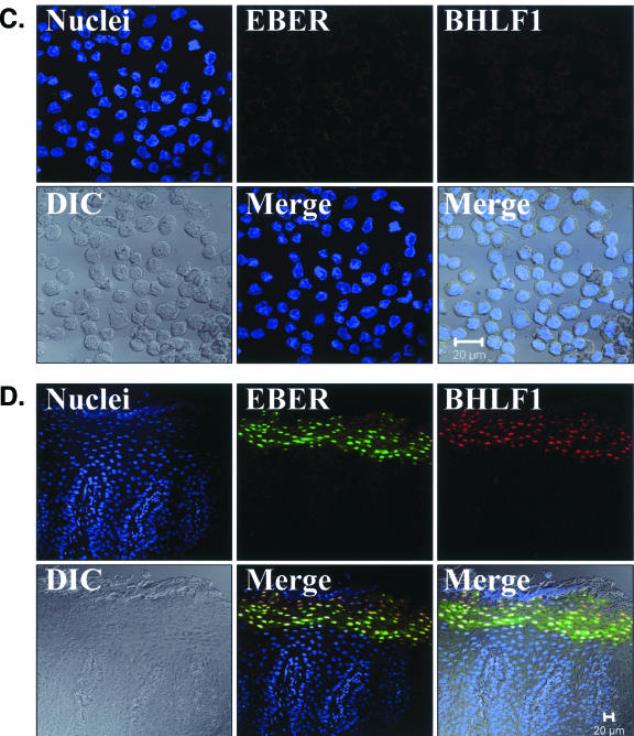
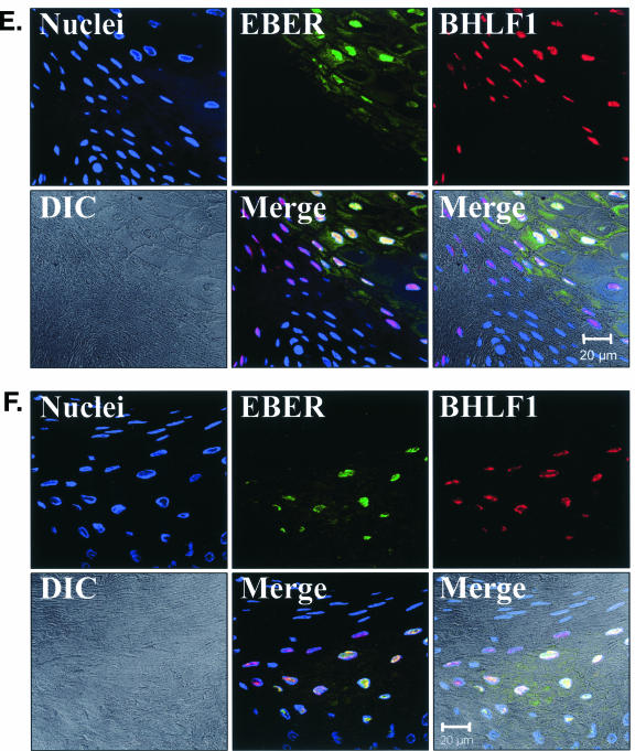
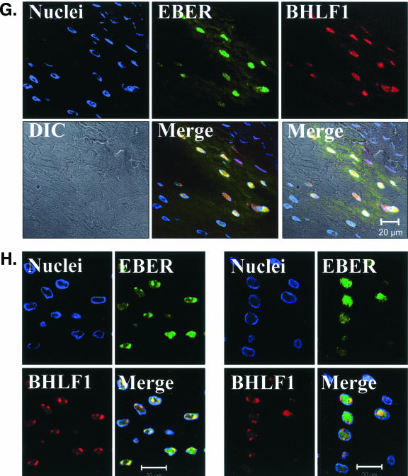
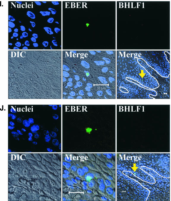
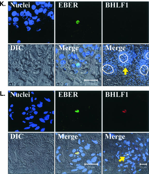
EBV EBER/BHLF1-FISH of oral epithelial tissue. Control cells and oral surgical biopsy tissue sections from HIV-positive subjects were examined by in situ hybridization for latency-associated EBER transcription and replication-associated BHLF1 transcription. Tissues were imaged by fluorescence laser scanning confocal microscopy. Tissue section panels are oriented with the mucosal surface to the top. DIC, Nomarski differential interference contrast. (A) EBV-positive B958 lymphoblastoid cells express high levels of EBER in most cells, and nuclear saturation of fluorescence was consistently detected. Approximately 1 to 5% of B958 cells also express replicative genes, including the early replicative gene BHLF1. Two different patterns of nuclear fluorescence were seen with BHLF1 expression, diffuse and punctate. (B) EBV-positive Namalwa Burkitt's lymphoma cells express much lower levels of EBER (at least 100-fold lower than B958 cells). Variable levels of nuclear EBER expression were easily detected in most Namalwa cells, but BHLF1 expression was not detected in these cells, which harbor only latent EBV infection. (C) EBV-negative RHEK-1 epithelial cells did not hybridize to either EBER or BHLF1. (D) Hybridization with both the EBER and BHLF1 probes was seen in a band-like pattern in the upper spinous layer of oral hairy leukoplakia, consistent with the known localization of productive EBV replication in the oral epithelium. (E, F, G, and H) Nuclear EBER probe hybridization was always associated with nuclear cohybridization of the BHLF1 probe in the upper spinous layer of oral hairy leukoplakia. Nuclear chromatin margination was present, and the most intense EBER probe hybridization strongly colocalized with the BHLF1 probe in the punctate hybridization pattern. This phenomenon of EBER probe hybridization in the upper spinous layer of oral hairy leukoplakia does not represent latent EBV infection but instead is consistent with EBER probe cross-hybridization to EBER gene sequences present in single-stranded EBV DNA synthesized in the nuclei of these cells during productive EBV replication, as previously described for EBER in situ hybridization in oral hairy leukoplakia (28). In the cells with the strongest nuclear EBER hybridization, additional weaker EBER hybridization was often seen in the cytoplasm and likely represents EBER probe cross-hybridization to EBER gene sequences present in artifactually denatured double-stranded EBV DNA in maturing virions being prepared for release from the cells. Furthermore, the cells immediately below and immediately above the EBER-BHLF1 cohybridizing cells often showed nuclear hybridization with only the BHLF1 probe. This result is consistent with early gene expression both preceding and persisting after viral DNA synthesis in the differentiation-dependent cascade of replicative EBV gene expression in oral epithelium, as previously described in oral hairy leukoplakia (40, 52). (I, J, and K) Three tissue sections (panel I, normal tongue epithelium without EBV replication; panels J and K, tongue epithelium with oral hairy leukoplakia) each demonstrated a solitary EBER-expressing cell located in or immediately above the basal layer. The locations of the epithelial basement membrane and basal layer are illustrated by the white lines and circles. The tissue section in panel K represents a cut through the mucosal rete ridges (white circles) in a plane that is perpendicular to the plane represented by the tissue sections in panels I and J. In all three cases, the EBER probe localization was confirmed to be intranuclear in a single cell by computer-generated three-dimensional reconstruction of the cell with a sequential series of 0.6-μm-deep confocal microscopy images. This expression of EBER in the absence of BHLF1 indicates the presence of latent EBV infection in each of these three solitary cells, similar to that demonstrated in the Namalwa cell line (panel B). (L) A tissue section of tongue epithelium with oral hairy leukoplakia demonstrated a solitary EBER- and BHLF1-coexpressing cell in the basal or lower spinous epithelial layer. The location of the epithelial basement membrane is not evident in this photomicrograph, but the cell appears to be located at the top of a rete ridge and was distinctly distant from the EBV replication in the upper spinous epithelial layer. The punctate nuclear colocalization of BHLF1 with the more diffuse EBER and the absence of EBER in the cell cytoplasm together suggest that this cell represents EBV reactivation of early replicative gene expression in a previously latently infected cell, similar to that demonstrated in the B958 cell line (panel A).

