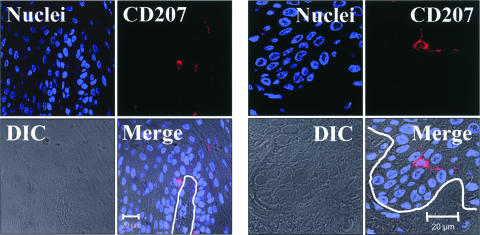
FIG. 7.
CD207/Langerin immunostaining of LC in oral epithelial tissue. Oral surgical biopsy tissue sections of normal tongue epithelium were immunostained for CD207/Langerin. Tissues were imaged by fluorescent laser scanning confocal microscopy. Tissue section panels are oriented with the mucosal surface to the top. DIC, Nomarski differential interference contrast. LC were identified in the basal layer and the immediate suprabasal region of the lower spinous layer of the oral epithelium. The location of the epithelial basement membrane and basal layer is illustrated by the white lines. These results demonstrate that oral LC localize to the same lower epithelial layers as the solitary EBV-positive cells identified in Fig. 6, suggesting a possible LC identity for these EBV-positive cells.

