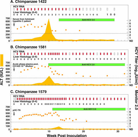
FIG. 1.
Course of infection with HCV strain HC-TN in (A) CH1422 (first-passage polyclonal infection), (B) CH1581 (second-passage quasipolyclonal infection), and (C) CH1579 (pHC-TN monoclonal infection). Serum samples collected once or twice weekly were tested for HCV RNA by an in-house RT-nested PCR with 5′ UTR primers and/or by use of a Roche Monitor 2.0 test. Red rectangle, positive by RT-nested PCR and/or by Monitor; white rectangle, negative by RT-nested PCR in two independent assays. The orange dots represent HCV Monitor titers; samples below the detection limit of 600 IU/ml (indicated by the dotted line) are shown as not detected (ND). Seroconversion in the second-generation ELISA is represented by a green horizontal bar. Yellow-shaded area, serum ALT. For liver histology, necroinflammatory changes of liver biopsy samples are graded 0 (normal), 1 (mild), 2 (mild to moderate), 3 (moderate to severe), or 4 (severe).

