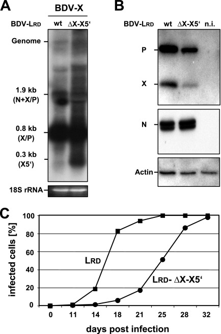
FIG. 2.
Comparison of viral gene expression and growth characteristics of BDV-LRD and BDV-LRD-ΔX-X5′ in Vero cells. (A) Northern blot analysis of RNA (samples of 20 μg) from Vero cells infected with BDV-LRD or BDV-LRD-ΔX-X5′. The blot was hybridized with a radiolabeled DNA probe specific for the BDV-X open reading frame. Note the additional 0.3-kb transcript originating from the ectopic X gene in cells infected with BDV-LRD-ΔX-X5′. (B) Western blot analysis of extracts (samples of 5 μg) from Vero cells that were either not infected (n.i.) or infected with BDV-LRD and BDV-LRD-ΔX-X5′. Blots were stained with antibodies specific for actin or the viral proteins P, X, and N. (C) Cultures infected with either virus at a multiplicity of 0.01 per cell were split twice weekly, and the proportion of infected cells was determined by immunostaining for the BDV N protein as described previously (6).

