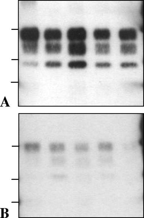
FIG. 3.
Western blot features of PrPres in TgOvPrP4 mice infected with, from the left to the right lanes in each panel, BSE, CH1641, TR316211, O104, and O100. In the last two cases, the samples were from one of the inoculated mice showing predominant l-type PrPres. PrPres was detected by Bar233 (A) and P4 (B) antibodies. The bars on the left of the panels indicate the 29.0-, 20.1-, and 14.3-kDa marker positions.

