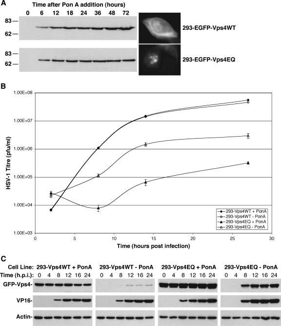
FIG. 2.
Single-step growth curves of HSV-1 in Vps4WT- and Vps4EQ-expressing cell lines. Clonal 293 cell lines expressing GFP-Vps4WT or GFP-Vps4EQ under the control of the ecdysone response element were isolated. (A) Cells were treated with 1 μM ponA for various times, and protein extracts were analyzed by Western blotting with a GFP-specific antibody. Fluorescence microscope images were collected at 16 h after ponA addition. Numbers at left are molecular masses in kilodaltons. (B) Cells were treated with or without ponA for 16 h and infected with HSV-1, and progeny virus was harvested at various times. Infectious viral titers were determined by plaque assay. Data represent mean PFU/ml, and error bars represent 1 standard deviation from the mean of triplicate samples. (C) Protein extracts were harvested from infected cells at various times postinfection and analyzed by Western blotting with GFP-, VP16-, and actin-specific antibodies.

