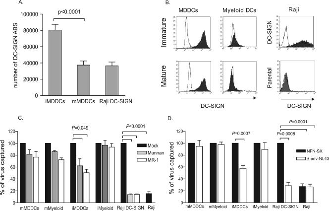
FIG. 4.
Higher viral capture in mature DCs than in immature DCs can occur independently of DC-SIGN and does not require viral envelope glycoprotein. (A) MDDCs of 30 seronegative donors were stained with anti-DC-SIGN mAb labeled with PE (1:1 ratio of mAb to PE) and analyzed for DC-SIGN surface expression using Quantibrite PE standard beads to calculate the number of DC-SIGN ABS displayed per PE-positive cell. Mean values and SEM are represented. iMDDCs had twice the mean number of DC-SIGN ABS per cell compared to those of mMDDCs and Raji DC-SIGN cells (P < 0.0001, paired t test). (B) Expression of DC-SIGN in MDDCs, myeloid DCs, Raji DC-SIGN cells, and Raji cells. Isotype-matched mouse IgG controls are also indicated (empty peaks). (C) Percentage of p24gag HIVNFN-SX captured at 37°C in the presence of different DC-SIGN inhibitors relative to untreated cells normalized to 100% of viral capture. Raji cells were compared to untreated Raji DC-SIGN cells. Viral capture by cells with no inhibitor (dark bars) or preincubated with mannan (light gray bars) or MR-1 (white bars) is depicted. Significant inhibition in the Raji DC-SIGN cell line reached 85% (P < 0.0001, paired t test). Mean p24gag (pg/ml) values of untreated cells used for normalization were 3,960 for mMDDCs, 1,154 for iMDDCs, 1,497 for mature myeloid cells (mMyeloid), 765 for immature myeloid cells (iMyeloid), 566 for Raji DC-SIGN cells, and 105 for Raji cells. Data show mean values and SEM from two independent experiments, including cells from six different donors. (D) Percentage of p24gag HIVΔenv-NL43 virus lacking the envelope glycoprotein captured by MDDCs, myeloid DCs, Raji cells, and Raji DC-SIGN cells relative to HIVNFN-SX capture normalized to 100%. Cells were pulsed with equal amounts of both viruses, and the Raji cell line was compared to Raji DC-SIGN cells. The envelope requirement for mMDDC and myeloid DC viral capture was not significant, while it reached significance in iMDDCs (P = 0.0007, paired t test). Raji DC-SIGN cells captured mainly HIVNFN-SX enveloped virus and bound HIVΔenv-NL43 virus only to levels comparable to the background seen by employing the Raji cell line (P = 0.0001, paired t test). Mean p24gag (pg/ml) values in cells exposed to HIVNFN-SX and used for normalization were 4,880 for mMDDCs, 1,562 for iMDDCs, 4,737 for mature myeloid cells, 1,280 for immature myeloid cells, 895 for Raji DC-SIGN cells, and 243 for Raji cells. Data show mean values and SEM from two independent experiments, including cells from five different donors.

