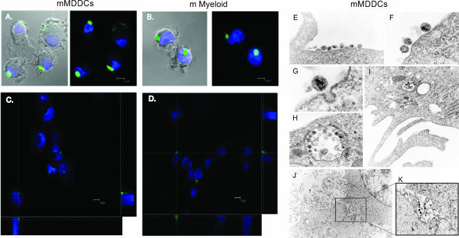
FIG. 5.
Confocal and electron microscopy of HIV1 capture and transfer mediated by mMDDCs. (A) Confocal images of a section of mMDDCs exposed to HIVNL43/eGFP for 4 h and stained with DAPI. Merged images of the section showing the cells and the green and blue fluorescence are shown. (B) Composite of a series of x-y sections collected through the entire thickness of the cell nucleus of mature myeloid DCs (m Myeloid) exposed to HIVNL43/eGFP and projected onto a two-dimensional plane. A three-dimensional reconstruction of a series of x-y sections collected through part of the cell nucleus can be found as movie S1 in the supplemental material. (C) Composition of a series of x-y sections of mMDDCs collected through part of the cell nucleus and projected onto a two-dimensional plane to show the x-z plane (bottom) and the y-z plane (right) at the points marked with the dotted white axes. (D) Confocal image composition of mature myeloid DCs constructed as described above (C). (E to G) mMDDCs presented large amounts of viral particles associated with the cell membrane outside dendrites (E and F) or initiating endocytosis (G). (H and I) Large vesicles containing numerous viral particles could be found proximal to the plasma membrane surface or deep inside the cellular cytoplasm. (J) Infectious synapse could also be observed in mMDDC and Hut CCR5+ cell cocultures. The marked box on the picture shows the attachment of mMDDCs (with light cytoplasm at the bottom) to a Hut CCR5+ cell (with granulated cytoplasm and a large nucleus at the top). (K) Magnification of the marked box in J is shown on the right, where viral particles are polarized to the cell-to-cell contact area, to form what has been described as a virological synapse.

