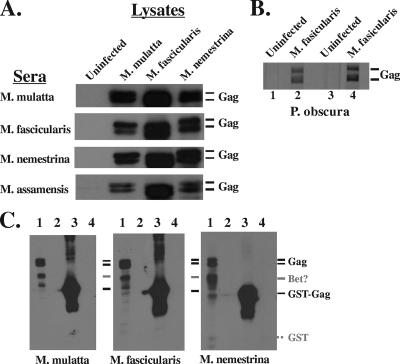
FIG. 3.
WB analysis of NHP sera reactivity with SFV Gag. Cell lysates were made from uninfected TF cells or cells infected with the indicated viruses. Viruses were from M. nemestrina and M. fascicularis, housed at WaNPRC; M. mulatta, housed at ONPRC; and M. assamenis and P. obscura, free ranging in Thailand. (A) The source of the lysates is shown at the top, and the source of the sera is on the left. Protein was detected using ECL reagents (Amersham). The lysates were not normalized for the amount of protein loaded; equal volumes were added to each lane. (B) Protein on individual membrane strips was detected using TMB. Lanes 1 and 2 used serum from one P. obscura animal, and lanes 3 and 4 used serum from a second animal. (C) WB were probed with sera from the indicated animals. Lanes 1, lysate from TF cells infected with SFV from M. fascicularis; lanes 2, lysate from uninfected TF cells; lanes 3, 1 μg of GST-Gag; lanes 4, 1 μg of GST. The dotted line indicates the position of GST not recognized by the sera.

