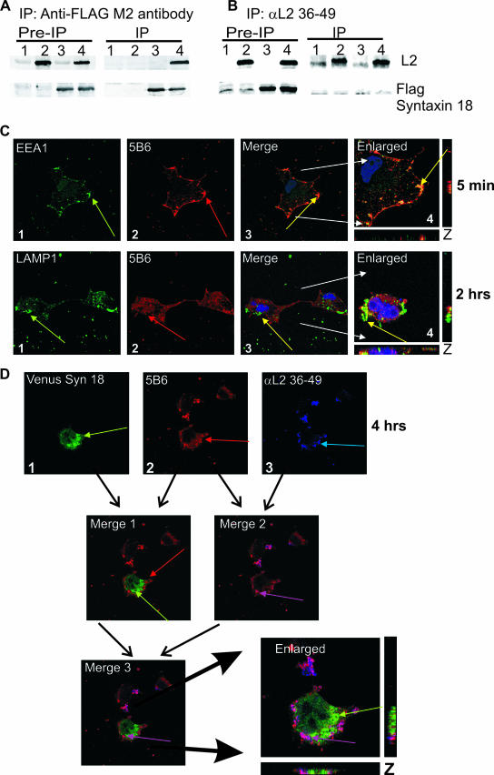
FIG. 8.
αL2 36-49 antibody interferes with the interactions of syntaxin 18 with L2 protein and wtL2 pseudovirions. (A and B) Cells were transfected with control vector pA3M (lane 1), pA3M and BPV1 L2 (lane 2), pA3M and FLAG-syntaxin 18 (lane 3), or BPV1 L2 and FLAG-syntaxin 18 (lane 4). Samples were immunoprecipitated (IP) with FLAG M2 antibody-coated beads (A) or αL2 36-49 (B). Western blots for L2 (top) and FLAG-syntaxin 18 (bottom) are shown. (C) αL2 36-49 antibody does not interfere with early viral entry. COS-7 cells were infected with wtL2 BPV1 pseudovirions preincubated with αL2 36-49 antibody for 5 min (top row) or 2 h (bottom row). Cells infected for 5 min (top row) were stained with anti-EEA1 (first panel, green arrow) and 5B6 (second panel, red arrow); cells infected for 2 h (bottom row) were stained with anti-LAMP1 (panel 1, green arrow) and 5B6 (panel 2, red arrow). Colocalization of the wtL2 pseudovirions and EEA1 or LAMP1 was not impeded by the presence of αL2 36-49 (top and bottom rows, panels 3 and 4, yellow arrows in the merged and enlarged images). (D) The addition of αL2 36-49 prevents the colocalization of BPV1 pseudovirions with syntaxin 18 at 4 h. Shown are VENUS syn 18 fluorescence (D1) (green arrow), BPV1 pseudovirions stained with 5B6 (D2) (red arrow), and staining of the αL2 36-49 pseudovirion-bound antibody (D3) (blue arrow). Merge 1 shows the overlay and lack of signal overlap of VENUS syn 18 and 5B6; merge 2 shows the overlap of signals for 5B6 and αL2 36-49, which results in a violet color (violet arrow); merge 3 and the enlarged image show the overlay of VENUS syn18, 5B6, and αL2 36-49 staining. The overlap of 5B6 and αL2 36-49 antibody staining (violet arrow) does not overlap with VENUS syn 18 (green arrow).

