
Figure 5.
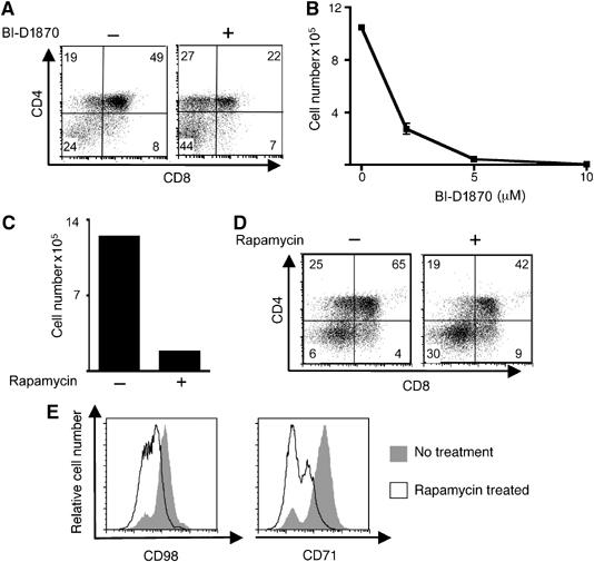
Inhibition of either mTOR or RSK permits thymocyte differentiation but not proliferation. (A) The data show surface expression of CD4 and CD8 on DN WT thymocytes following 3 days co-culture with 5 ng/ml IL7 on OP9-DL1 monolayers in the presence or absence of 10 μM BI-D1870. The data are representative of two independent experiments. (B) Data show numbers of cells following 6 days co-culture of DN thymocytes on OP9-DL1 in the presence of BI-D1870 at concentrations indicated. (C) Data show the number of cells following 6 days co-culture of DN thymocytes co-cultured on OP9-DL1 monolayers with 5 ng/ml IL7 in the presence or absence of 20 nM rapamycin. Data are representative of three independent experiments. (D) Data show CD4 and CD8 expression on DN3 WT thymocytes following 6 days co-culture with 5 ng/ml IL7 on OP9-DL1 in the presence or absence of 20 nM rapamycin. Data are representative of three independent experiments. (E) Data show CD98 (left panel) and CD71 (right panel) surface expression on DN3 thymocytes, following 2 days co-culture with 20 nM rapamycin on OP9-DL1. Data are representative of three independent experiments.