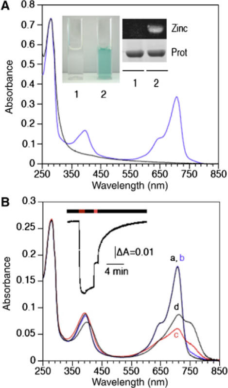
Figure 2.

Absorption spectra of RpBphP4 proteins. (A) Absorption spectra of recombinant RpBphP4 proteins from strains CEA001 (achromo-RpBphP4, black line) and HaA2 (chromo-RpBphP4, blue line). The inset shows (1) achromo- and (2) chromo-RpBphP4 proteins. The covalent binding of BV to RpBphP4 proteins was visualized by zinc-induced fluorescence or Coomassie staining. (B) Absorption spectra of purified HaA2 RpBphP4. The spectra were recorded immediately after: 770 nm illumination (a, black line) followed by 15 min of darkness (b, blue line), 705 nm illumination (c, red line) followed by 15 min of darkness (d, black line). The inset shows the kinetics of changes in absorbance at 750 nm induced by 770 nm light, followed by 2 min of darkness, 1 min of 705 nm light and then a long dark period.
