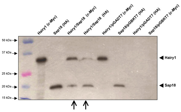
Figure 3.
In vitro co-immunoprecipitation of Hairy1 and Sap18. The proteins tested are indicated on top of each lane, as well as the antibody used for immunoprecipitation in each assay (in brackets). Proteins produced form the pGADT7 and pGBKT7 empty plasmids were used in negative controls. Open arrows indicate the gel lanes where both Hairy1 and Sap18 proteins are observed, which is indicative of a protein interaction among them.

