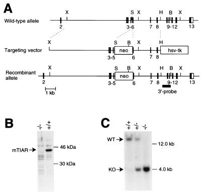
Figure 1.
Gene targeting at the tiar locus. (A) Schematic representation of the tiar gene exon–intron structure with restriction enzyme sites (11), the structure of the targeting vector, and the structure of the tiar locus after integration of the targeting vector. Correct gene targeting results in replacement of portions of intron 5 and exon 6 by the marker gene for positive selection (PGKneo). The hsv-TK (thymidine kinase) expression cassette was used for negative selection. The 3′ probe used for Southern blot analysis is shown as a stippled box. B, BamHI; H, HindIII; S, SalI; X, XbaI. (B) Protein immunoblot of total lysates of tiar−/− and tiar+/+ ES cells by using the anti-TIAR mAb 6E3 (11) confirming absence of TIAR protein in tiar−/− cells relative to wild-type cells. Use of an anti-TIAR mAb reactive with a different TIAR epitope (anti-3E6) (11) gave the same result (data not shown). (C) Southern blot analysis of DNA from offspring derived from heterozygous matings. Genomic DNA was digested with BamHI and analyzed by using the 3′ probe, yielding the >12-kb and 4.0-kb fragments expected for the wild-type allele and mutant allele, respectively. Southern blot analysis of ES cell DNA using a 5′ DNA probe and a neo DNA probe showed proper targeting and single insertion of the transfected vector (data not shown).

