
Figure 6.
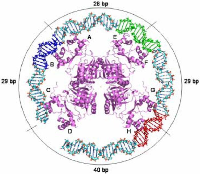
Model of the E. coli Lrp octamer bound to Lrp binding sites 1, 2, and 3 of the pap reguglatory region. Lrp binding site 3 is colored red, site 2 is depicted in green, and site 1 is shown in blue. The labels on the periphery indicate the number of base-pairs that separate the center two-fold axes of the individual dimers from each other.