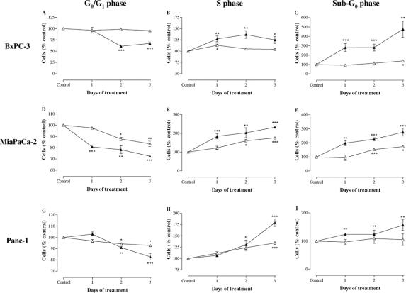
FIGURE 4. Cell cycle distribution after 1, 2, and 3 days of incubation with 1000 IU/mL IFN-α and 1000 IU/mL IFN-β in BxPC-3 (A–C), MiaPaCa-2 (D–F), and Panc-1 (G–I) cells. Data are expressed as mean ± SEM of the percentage of cells in the different phases of the cell cycle, as compared with untreated control cells. Control values have been set to 100%. Δ, IFN-α; ▴, IFN-β. *P < 0.05; **P < 0.01; ***P < 0.001 versus control.

An official website of the United States government
Here's how you know
Official websites use .gov
A
.gov website belongs to an official
government organization in the United States.
Secure .gov websites use HTTPS
A lock (
) or https:// means you've safely
connected to the .gov website. Share sensitive
information only on official, secure websites.
