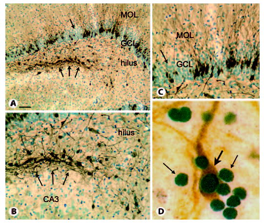
Fig. 2.

Calbindin and PROX1 expression in the epileptic rat. MOL = Molecular layer; GCL = granule cell layer. A Calbindin and cresyl violet staining of a section through the septal pole of a pilocarpine-treated rat with recurrent seizures illustrates irregular loss of calbindin expression despite persistence of apparently normal granule cell cresyl violet staining (arrow) and clusters of calbindin-immunoreactive cells at the border of the hilus and CA3 region (triple arrows). Calibration = 100 μm. B Higher magnification of the clusters of hilar calbindin cells shown in A. Calibration (in A) = 50 μm. C Higher magnification of the irregular loss of calbindin in the granule cell layer (arrow in A). Calibration as for B. D Double-labeling of sections from the hilus of a pilocarpine-treated rat with recurrent seizures demonstrates that numerous PROX1-immunoreactive nuclei are present (small arrows), but only one is double-labeled with calbindin (orange, large arrow), suggesting that PROX1 is the more reliable marker of mature granule cells than calbindin in rats with chronic seizures. Calibration (in A) = 5 μm.
