
Fig. 4.
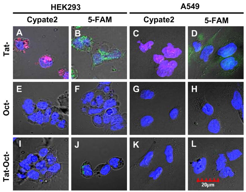
Intracellular distribution of molecular probes (1 μM) in HEK293 and A549 cells at 37 ºC for 30 min. Incubation of Tat-cypate2, Oct-cypate2, and Tat-Oct-cypate2 (A, E, I) or Tat-5-FAM, Oct-5-FAM, and Tat-Oct-5-FAM (B,F,J) with HEK cells and Tat-cypate2, Oct-cypate2, and Tat-Oct-cypate2 (C, G, K) or Tat-5-FAM, Oct-5-FAM, and Tat-Oct-5-FAM (D, H, L) with A549 cells. Color scheme: nuclear stain, blue; 5-FAM, green; Cypate2, red; Green/blue overlap, light green; red/blue overlap, purple.