
Figure 1.
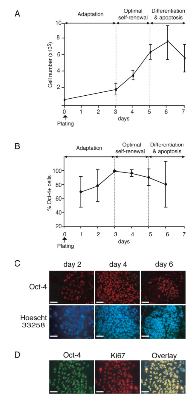
Population doubling time and expression of Oct-4 and Ki67 markers in ORMES-1 cells. ORMES cells were plated on inactivated MEF at a density of 0.3 × 10⁵ cells per cm2. (A) Cell numbers were counted at each time point (2 to 3 replicates) in two independent experiments using a Trypan blue exclusion assay. Means and standard errors to the mean (SEM) are indicated on the graph. (B) Immunohistofluorescent detection of Oct-4 (revealed by Cy3) in undifferentiated ORMES-1 cells between day 1 and day 6 of culture. Curve represents the mean percentages of Oct-4+ cells within colonies (n = 7 to 14) of ORMES-1 cells. (C) Immunohistofluorescent detection of Oct-4 (revealed by Cy3) in ORMES-1 cells at day 2, 4 and 6 of the culture (D) Immunohistofluorescent detection of Oct-4 (revealed by Cy2) and Ki-67 (revealed by phycoerythrine) in ORMES-1 cells at day 3 of the culture. (C,D) Bar = 10 μM.