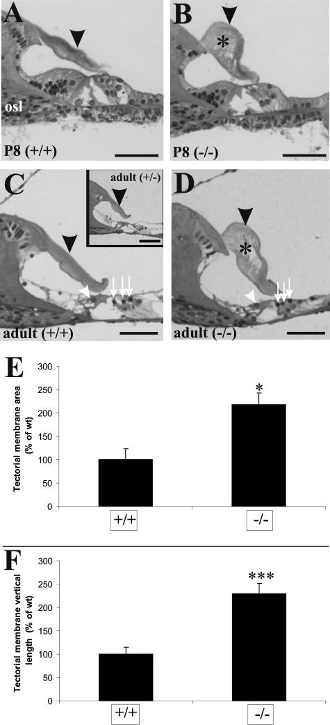
Figure 2.
Tectorial membrane defects in Tmprss1−/− mice. Histological analysis of cochleae from wild-type and Tmprss1−/− mice at postnatal day 8 (P8) (A and B, respectively) and adult (C and D, respectively) mice. Black arrowheads point to the tectorial membrane, and asterisks show holes found within the matrix of the membrane. Osseous spiral lamina is indicated by osl. White arrowheads show inner hair cells, and white arrows point to the outer hair cells in the organ of Corti. Scale bar = 50 μm. C, inset: Analysis of cochleae from adult Tmprss1+/− mice. E: Comparison of the cross-sectional area of the tectorial membrane in wild-type and Tmprss1−/− mice. Graph indicates mean ± SE, *P = 0.011. F: Comparison of the longest vertical distance measured between the dorsal and ventral surface of the tectorial membrane. Graph indicates mean ± SE, ***P = 0.002.

