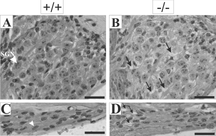
Figure 3.
Morphological defects in the Rosenthal’s canal and osseous spiral lamina of Tmprss1−/− mice. Representative images of the Rosenthal’s canal and the osseous spiral lamina of adult wild-type (A and C, respectively) and adult Tmprss1−/− (B and D, respectively) mice. A: White arrowhead indicates spiral ganglion neuron (SGN). B: Black arrows indicate spaces between spiral ganglion neurons. C: White arrowhead indicates spindle-shaped nuclei of Schwann cells. D: Black arrows point to spaces between Schwann cells. Scale bar = 20 μm.

